- 13a
- 13b
- 13c
- 13d
- 13e
Figures
Fig. 13a: Patient with scull base tumor after 2 operation (via antral and neurosurgical approach), with defect of infraorbital region and orbital floor, enophthalmos, ptosis of upper eyelid, paralysis of VI nerve and blindness.


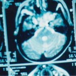
Fig. 13b: MRI scan of scull base tumor.


Fig. 13c: Design of incision.


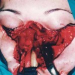
Fig. 13d: Le Fort III osteotomy of facial bones and sagital separation. By this approach it was possible to remove scull base tumor.


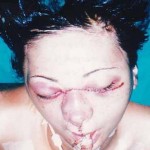
Fig. 13e: Finish of operation. All tissue are moved in original position.
« back to Tumors of Craniofacial Region


