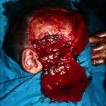
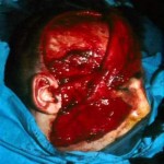
- 3a
- 3b
- 3c
- 3d
Figures
Fig. 3a: Preoperative view


Fig. 3b: The same patient 3 weeks postoperatively. Correction of facial deformity is performed using pericranial flap pedicled on temporal vessels.


Fig. 3c: The pericranial flap is raised.


Fig. 3d: The pericranial flap is doubled and placed over the skin to simulate the future position in the subcutaneous pocket.
« back to Facial Deformities


