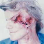
- 14a
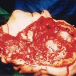
- 14b
Figures
Fig. 14a: The patient with basal cell carcinoma which involved orbit, scull base, maxilla, frontal, temporal, occipital bone, mandible and ear.


Fig. 14b: Intraoperative view

Frontal, temporal, part of occipital bone, orbit, maxilla, zygoma, ramus of mandible, part of scull base and ear are removed. Cervical vessels are prepared for microsurgical anastomosis of myocutaneous latisumus dorsi flap.
« back to Tumors of Craniofacial Region


