- 9a
- 9b
- 9c
Figures
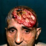
Fig. 9a: Preoperative view


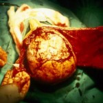
Fig. 9b: Intraoperative view - tumor is removed with affected bone and two large scalp flaps are raised in order to cover defect of scalp and bone in frontal region.


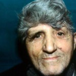
Fig. 9c: The same patient one year after operation.
« back to Tumors of Craniofacial Region


