- 30a
- 30b
- 30c
- 30d
Figures
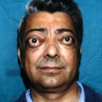
Fig. 30a: Preoperative view


Fig. 30b: Patient two month after 3-walls orbital decompression, removal of orbital fat and correction of the eyelid deformities.


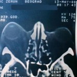
Fig. 30c: Preoperative CT scan


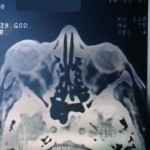
Fig. 30d: Postoperative CT scan
« back to Tumors of the Orbit and Thyroid Ophthalmopathy


