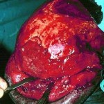
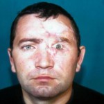
- 25a
- 25b
- 25c
Figures
Fig. 25a: Preoperative view


Fig. 25b: Osteopericranial flap is raised on pericranium, i.e. on temporal vassels and placed in recipient region.


Fig. 25c: Postoperative view after first operation - defect of fronto-orbital region is corrected using vascularised calvariar osteopericranial flap pedicled on the temporal vessels.
« back to Injuries of Craniofacial Region


